IMPLANTABLE LOOP RECORDER
This is a type of heart rhythm monitoring device, the size of a standard pen drive. It is placed under the skin of your chest by a minor procedure. This records the electrical activities of your heart continuously and can remain in place for up to 3 years. We get valuable information about abnormal (fast or slow) heart rhythms which could be responsible for your symptoms. This device can be interrogated without taking it out in the event of you having a fainting episode.

EP STUDY
We map the electrical activity within your heart to identify the exact cause of abnormal rhythms. EP studies help determine which treatment will be effective for you and in certain situations can predict the risk of sudden cardiac death. We will place diagnostic catheters within your heart and run specialized tests to map the electrical currents. This carries a small risk of complications. In certain situations, your EP study may be done via catheters placed in the food pipe. This is called a transesophageal EP study.

CORONARY ANGIOGRAM
This is a procedure that uses X-rays to see your heart's blood vessels and identify any restriction in blood flow to the heart. During this test, we will insert a flexible tube (catheter) through your groin or arm to the heart. A type of dye that's visible by an X-ray machine is then injected into the blood vessels of the heart and this produces images of the blood vessels.

TRANSESOPHAGEAL ECHO (TEE)
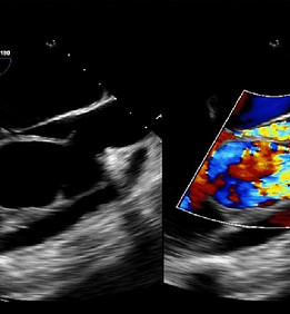
We may sometimes need more detailed images of the heart than what can be produced by the standard transthoracic ECHO. TEE is used in such situations to visualize the heart. A flexible tube is inserted via the mouth into the food pipe. This probe will transmit pictures of your heart to the computer. You will be given medications to numb your throat before the procedure to minimize your discomfort. You will be allowed to have clear liquids 2 hours after this test and solids four hours later.

INTRA CARDIAC ECHOGRAPHY
We may have to perform Intracardiac Echography (ICE) during your radiofrequency ablation procedure. It aids us in complex transseptal punctures, baffle punctures and to help position the ablation catheter over the papillary muscle in papillary muscle ventricular tachycardia. The ICE catheter is inserted through a vein in the neck or the groin into the heart. It then transmits pictures through a fiber optic cable to be seen on a computer screen.

ESOPHAGEAL EP STUDY
This test is done in children less than 10 years of age. The test involves placing a bipolar or quadripolar lead through the nostril into the oesophagus. It is most often performed in new-born or babies less than 2 years to assess the presence of a tachyarrhythmia and to help evaluate if it is well controlled on medication. It has a therapeutic use in children with sinus node dysfunction by aiding with atrial pacing.

